Fall 2025 - Vol. 20, No. 3
PHOTO QUIZ FROM APP ONBOARDING PROGRAM
A Handful of Trouble
Casey Hershey, MSN, CRNP, FNP-C
Nurse Practitioner, Family Medicine Queen Street
Penn Medicine Lancaster General Health Physicians
CASE HISTORY
A 66-year-old female presents to the office with complaints of a sore on her left dorsal hand that initially presented 10 days ago. She first noticed the sore after scraping her hand against a car seat resulting in mild swelling and erythema; she did not experience skin breakdown (see Fig. 1). The patient reports it feels like a foreign object is in her hand.
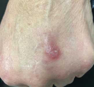
Fig. 1. Patient’s sore, day 10, as first presented in office.
She has already tried lancing it herself at home, after which a small amount of old blood was released, but it remains painful. The left hand has what appears to be an abscess that is mobile, soft, round, and erythematous, measuring approximately 1 x 1 cm in size; it is not warm to the touch.
The patient denies any fevers, myalgias, or chills. Concurrently, she began Keflex® for group A streptococcal pharyngitis two days prior to presenting to the office. She further relays that she actively swims in a chlorinated pool two days a week and takes Plaquenil® and methotrexate for rheumatoid arthritis. It is recommended that she continue Keflex® and topical Bactroban™ for suspected cellulitis with potential abscess.
The patient returns a week later with no improvement on Keflex® and Bactroban™. The wound/abscess on her left dorsal hand is erythematous, tender, and warm to touch; it measures 1.8 x 1 cm in size with a new 1 cm induration noted at the wound bed (see Fig. 2).
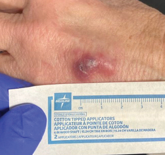
Fig. 2. Patient’s sore at second visit, a week later, showing no improvement on Keflex® and Bactroban™.
At this office visit, an incision and drainage of the abscess on her left dorsal hand is performed, yielding purple-yellow pus. A wound culture is obtained, and she is started on doxycycline in addition to the Keflex®.
The patient presents back in the office two weeks later with no improvement in her left-hand abscess after finishing a seven-day course of doxycycline. Her left hand has an erythematous maculopapular lesion measuring approximately 1.8 x 2 cm in size that is not draining but is tender (see Fig. 3).
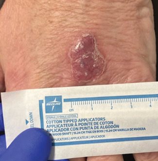
Fig. 3. Patient’s sore, two weeks post-drainage, showing no improvement after a seven-day course of doxycycline.
An e-consult with Infectious Diseases is initiated after which the patient is prescribed rifampin 300 mg oral daily and clarithromycin 500 mg oral two times a day for six weeks.
Three weeks later the patient calls into the office reporting the abscess is not any smaller, although it is also not as red, swollen, or tender. It is not responding to rifampin and clarithromycin and is now persistently draining purulent fluid. Exam of the patient’s left dorsal hand reveals a tender, round, erythematous/violaceous, maculopapular lesion that measures approximately 2 x 2.5 cm in size with a small open area draining a scant amount of serosanguinous fluid (see Fig. 4).
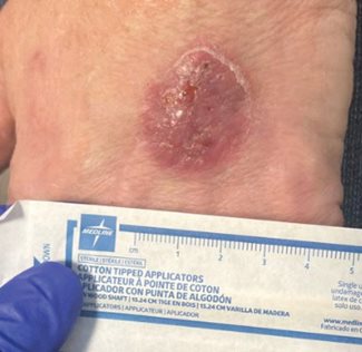
Fig. 4. Patient’s sore, five weeks post-drainage, showing no improvement on rifampin and clarithromycin.
At this visit, she is instructed to continue rifampin and clarithromycin therapy for three more weeks; an urgent referral is placed to Dermatology for potential biopsy and further recommendations.
That same week, the patient is seen by Dermatology for tangential biopsy. She later messages into the office to report her wound is constantly draining from the biopsy site.
QUESTIONS
- What is the differential diagnosis for this patient?
- What are the signs and symptoms of cellulitis?
- Who is at risk for cellulitis?
- What are the most common microbial pathogens in cellulitis?
- What is the difference between cellulitis and an abscess?
- How does the treatment differ between cellulitis and an abscess?
ANSWERS
- Potential diagnoses include but are not limited to cellulitis of the dorsal hand, simple cutaneous abscess, infected insect/animal bite, retained foreign body, ruptured/infected epidermal inclusion cyst, contact dermatitis, atopic dermatitis, tinea corporis, and squamous cell carcinoma.
- Symptoms of cellulitis include erythema, edema, warmth, and pain, and patients may present with purulent drainage and fever.
- Most middle-aged and older adults who experience a disruption in their skin barrier because of injury are at higher risk of developing cellulitis. Patients who have a history of obesity, eczema, psoriasis, venous insufficiency, immunosuppression, diabetes, and/or pre-existing skin infection are at higher risk of developing cellulitis if there is a break in the skin barrier.
- The most common pathogens in cellulitis are beta-hemolytic streptococci (groups A, B, C, G, and F), Streptococcus pyogenes, Staphylococcus aureus, and methicillin-resistant Staphylococcus aureus (MRSA).
- Patients with cellulitis may or may not present with an abscess. A skin abscess is a collection of pus that is fluctuant, often with an erythematous nodule.
- Patients who present with cellulitis should be started on antibiotics that will cover the suspected pathogen. Initial antibiotics for cellulitis should cover beta-hemolytic streptococci. If a patient presents with purulent wound drainage, toxic symptoms (fever >100.5°F, hypotension, tachycardia), or has recently been hospitalized or resides in a long-term care facility, antibiotics for MRSA coverage may be warranted. Patients who present with an abscess should undergo incision and drainage. After draining an abscess, antibiotics are not typically warranted because incision and drainage is the definitive treatment; however, if the patient is experiencing severe local infection or systemic symptoms, fails to respond to initial antibiotic, or has experienced an animal bite, they may be started on antibiotics after undergoing an incision and drainage. Additionally, patients who are immunocompromised or extremely young or old should be started on antibiotics after an incision and drainage.
ADDITIONAL CASE HISTORY
The wound culture grows
Tsukamurella tyrosinosolvens but the lesion does not respond to doxycycline, rifampin, or clarithromycin. Biopsy results show giant cells, and staining is positive for herpes simplex virus 1 and 2. The immunostains suggest an old herpes virus infection, and thus the patient is started on Valtrex™ and minocycline by her dermatologist. Minocycline is shortly discontinued by her Infectious Diseases specialist due to low suspicion that this is a bacterial infection, and Valtrex™ is increased to 1 gram every eight hours for two weeks.
Two weeks later, the patient reports her left-hand lesion is finally starting to improve (see Fig. 5). If the wound had not improved after the additional week of Valtrex™, minocycline could have been added.
 Fig. 5. Patient’s sore showing improvement, eight weeks after initial visit and two weeks after initiation of Valtrex™.
DISCUSSION
Fig. 5. Patient’s sore showing improvement, eight weeks after initial visit and two weeks after initiation of Valtrex™.
DISCUSSION
Herpes simplex should be considered in any case of a new painful cutaneous lesion, especially those that leak pustular fluid. Testing can include Tzank smear, but polymerase chain reaction testing is more readily available. Treatment with acyclovir or valacyclovir as early in the course of disease as possible is recommended for 10 days and possibly longer depending on reassessment.
Tsukamurella tyrosinosolvens is a rare Gram-positive acid-fast bacillus that belongs to the class Actinomycetes. These bacteria are found in soil and water and may be an opportunistic pathogen that particularly affects immunocompromised individuals and those with indwelling medical devices (peripherally inserted central catheter lines, cardiac pacemaker implants, etc.).
1
Tsukamurella are very similar to other species such as
Rhodococcus, Goronia, Corynebacterium, Nocardia, and
Mycobacterium.2,3 The most effective treatment strategy is starting appropriate antibiotics quickly and, if it is thought to be a device-associated infection, removing the indwelling medical device during antibiotic therapy.
2
REFERENCES
1. Aflatooni S, Kucharik AH, Fourzali KM, Turner L, Kowalewski C. Cutaneous
Tsukamurella tyrosinosolvens infection in an immunocompetent patient.
JAAD Case Rep. 2024;49:44-46.
2. Usuda D, Tanaka R, Suzuki M, et al. Obligate aerobic, Gram-positive, weak acid-fast, nonmotile bacilli,
Tsukamurella tyrosinosolvens: minireview of a rare opportunistic pathogen.
World J Clin Cases. 2022;10(24):8443-8449.
3. Liu CY, Lai CC, Lee MR, et al. Clinical characteristics of infections caused by Tsukamurella spp. and antimicrobial susceptibilities of the isolates.
Int J Antimicrob Agents. 2011;38(6):534-537.