Fall 2026 - Vol. 21, No. 3
CLINICAL REPORT
Mucocutaneous Changes in a Nine-year-old Boy
Karousatos Shumaker Durham
Alec Karousatos
Medical Student, Lewis Katz School of Medicine
Kimberly Shumaker, MD
Resident in Family Medicine
Penn Medicine Lancaster General Health
Megan Durham, MD
Assistant Medical Director, Hospital Pediatrics
Penn Medicine Lancaster General Health
PRESENTATION
A 9-year-old boy presents to a community hospital with mucocutaneous changes following a lower respiratory tract illness. One week prior, he developed a fever, cough and congestion, and was diagnosed with left lower-lobe community-acquired pneumonia. He began empiric treatment with amoxicillin at the onset of symptoms, but after five doses, the patient developed episodes of circumoral edema and mucosal ulceration of the mouth and lips.
Concerned for a possible allergic reaction, the patient’s primary care physician transitioned him from amoxicillin to azithromycin and started a course of oral corticosteroids four days ago. His parents report that he had previously tolerated amoxicillin without any adverse effects. His fever initially resolved, and his cough and congestion improved on antibiotics. However, his fever returned today accompanied by worsening mucocutaneous involvement (see Fig. 1) and new onset bilateral periorbital edema.
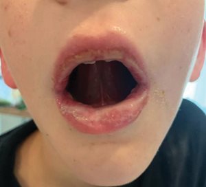
Fig. 1. Oral mucosal involvement on admission.
He reports intermittent blurred vision but denies pain with eye movements. His oral intake is poor, and he speaks sparingly due to pain.
On examination, the boy is ill-appearing but non-toxic and in no acute distress. His temperature is 100.1°F, pulse 90 bpm, respiratory rate 20 breaths/min, and blood pressure 105/60 mmHg. His pulse oximetry on room air is 100%. His mucous membranes are dry. He exhibits circumoral edema, labial mucosal skin sloughing, and ulcerations of the gingival and buccal mucosa. Bilateral periorbital erythema and edema are present, with crusted, purulent ocular discharge. Bilateral sub-centimeter anterior cervical lymph nodes are palpable. His capillary refill time is 2 to 3 seconds. There is no genital involvement.
A complete blood count shows no leukocytosis or neutrophilic predominance. A comprehensive metabolic panel reveals normal electrolytes, transaminases, and kidney function. Diagnostic testing on admission includes a negative respiratory viral panel, negative mononucleosis screening, and negative
Streptococcus pyogenes (group A) test. A repeat chest radiograph shows improvement in the recent left lower-lobe pneumonia. Physical exam findings and additional laboratory testing confirm the diagnosis.
DISCUSSION
Differential Diagnosis
The differential diagnosis for mucocutaneous changes in a child following an upper respiratory infection treated with antibiotics includes Stevens-Johnson syndrome (SJS); toxic epidermal necrolysis (TEN); erythema multiforme; reactive infectious mucocutaneous eruption (RIME); Kawasaki disease; herpes-simplex gingivostomatitis; paraneoplastic pemphigus; aphthous stomatitis; and hand, foot, and mouth disease (HFMD).
Actual Diagnosis
Serum
Mycoplasma pneumoniae titers demonstrate an IgG >5.00 U/L, indicative of a recent infection when correlated with chest X-ray findings and clinical presentation. The patient’s lesions are most prominent on the mucosal surfaces of the mouth, and the lack of cutaneous findings means that the diagnosis of SJS, TEN, or erythema multiforme are unlikely.
While both herpes-simplex gingivostomatitis and HFMD can cause oral ulcers in children, these diseases do not typically produce the extensive, bilateral periorbital erythema and edema seen in this child. In HFMD, additional vesicular lesions of the extremities would be expected as well. Given the presence of oral mucosa ulceration, conjunctivitis, and minimal cutaneous involvement in the setting of a
M. pneumoniae infection, a diagnosis of RIME is established.
The Condition
RIME is an immune-mediated mucocutaneous disorder primarily affecting children and adolescents following a bacterial or viral respiratory infection, most commonly
M. pneumoniae.1 Although the exact pathogenesis remains unclear, RIME is thought to arise either indirectly from immune complex formation against infectious antigens
2 or directly from local inflammatory cytokine release.
3
It is characterized by prominent mucositis, often involving the oral, ocular, and genital mucosa, with minimal or absent cutaneous involvement.
4 Patients typically present with a prodromal illness, including fever, cough, and malaise, followed by the development of painful mucosal ulcerations and conjunctivitis.
Until 2015, RIME was thought to be a viral-induced SJS but has since been differentiated.
2 In comparison to SJS or TEN, RIME has more prominent mucosal involvement and lacks extensive cutaneous findings.
5 If cutaneous lesions are present, they often present as scattered, atypical targetoid lesions.
4
Diagnosis is clinical, supported by confirmatory laboratory testing for suspected infectious agents. Most cases of RIME will resolve completely with supportive care; however, recurrent episodes have been reported.
6 Early recognition and management are crucial to preventing complications, particularly those involving the eyes.
Treatment/Management
The management of RIME is primarily supportive, including symptom relief, mucosal healing, and prevention of complications. Mucositis can be painful and severely impact oral intake; therefore, it is crucial to provide adequate pain control, intravenous fluids, and, in severe cases, nasogastric feeding. Pain control regimens often include acetaminophen and nonsteroidal anti-inflammatory drugs (NSAIDs), with opioids reserved for severe cases. Topical therapies such as petroleum jelly for the lips and anesthetic mouthwashes can provide additional relief.
Systemic corticosteroids should be considered in severe cases with extensive mucosal involvement. Prednisone and intravenous methylprednisolone are frequently used to reduce inflammation and expedite symptom resolution.
4 Other immunomodulatory agents, such as cyclosporine, intravenous immunoglobulin, or tumor necrosis factor inhibitors (e.g., etanercept), have been explored in refractory cases, with some evidence suggesting potential benefit.
7-9
Antimicrobial therapy for community-acquired pneumonia commonly includes macrolides, fluoroquinolones, beta-lactams, or tetracyclines (for children over 8 years old) depending on the identified pathogen. In cases of suspected
M. pneumoniae infection, empiric treatment with antibiotics is recommended, although there is not sufficient evidence to support that treatment will shorten the course of RIME.
10
Ophthalmology consultation should be obtained for any patient with ocular symptoms. To prevent long-term ophthalmologic complications, patients often receive frequent saline rinses, artificial tears, and topical corticosteroids (e.g., prednisolone ophthalmic drops); however, additional interventions may be required in cases of severe ocular disease.
11
PATIENT COURSE
By the time of admission, the patient had completed a five-day course of azithromycin and a four-day course of oral corticosteroids without improvement. Due to reduced oral intake, he receives maintenance intravenous fluids with 5% dextrose in 0.9% normal saline. Supportive care includes petroleum jelly for lip protection and scheduled intravenous acetaminophen with as-needed ketorolac for pain management.
On the second day of admission, he begins experiencing worsening mucosal lesions of the lips (see Fig. 2), increased pain, and decreased visual acuity.
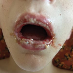
Fig. 2. Worsening oral mucosal involvement noted two days after photo in Fig. 1 was taken.
Pediatric dermatology and ophthalmology are consulted by telephone, recommending subcutaneous etanercept and tobramycin/dexamethasone ophthalmic ointment. Due to etanercept unavailability at the community hospital, he is transferred to a facility with pediatric subspecialists available for further management.
At the receiving hospital, ophthalmologic examination reveals mucus discharge with crusting of the lashes in both eyes. The conjunctivae are white and quiet, with no staining on the cornea and no evidence of epithelial sloughing or pseudo-membranes. Given that the patient lacks severe eye involvement and has already been symptomatic for a few days, etanercept is not initiated. Instead, he is given a three-day course of intravenous methylprednisolone, along with prednisolone ophthalmic drops and artificial tears. Morphine is made available for severe pain.
The patient’s condition improves with this treatment. He is able to demonstrate consistent oral fluid intake and therefore is deemed appropriate for discharge. At this time, all ophthalmic drops are discontinued, and supportive care with petroleum jelly and anesthetic mouthwashes are recommended for persistent mouth pain.
He continues to experience pain due to oral mucosal ulcerations while at home for another week before he improves enough to eat solid foods. At his ophthalmology follow-up appointment two weeks later, his symptoms have resolved.
LESSONS FOR THE CLINICIAN
- RIME causes mucositis in children most commonly following an infection with M. pneumoniae.
- RIME should be included in the differential diagnosis when a child presents with mucosal ulceration, conjunctival involvement, and a recent upper respiratory illness, particularly in the absence of widespread cutaneous involvement.
- Unlike Stevens-Johnson syndrome or toxic epidermal necrolysis, RIME is strongly associated with infectious triggers rather than medication exposure and typically lacks extensive skin involvement.
- Pain control, hydration, and nutritional support are key aspects of management, as oral mucosa involvement can significantly impair intake.
- Early consultation with ophthalmology is critical to prevent long-term complications such as scarring and vision loss.
- Systemic corticosteroids may be useful in treating the mucosal lesions of the disease. Immunomodulatory agents may be considered for severe or refractory cases when standard therapies fail to adequately control worsening mucosal lesions.
REFERENCES
1. Meyer Sauteur PM, Theiler M, Buettcher M, Seiler M, Weibel L, Berger C. Frequency and clinical presentation of mucocutaneous disease due to mycoplasma pneumoniae infection in children with community-acquired pneumonia.
JAMA Dermatol. 2020;156(2):144-150.
2. Simecka JW, Ross SE, Cassell GH, Davis JK. Interactions of mycoplasmas with B cells: antibody production and nonspecific effects.
Clin Infect Dis. 1993;17(Suppl 1):S176-S182.
3. Stutman HR. Stevens-Johnson syndrome and Mycoplasma pneumoniae: evidence for cutaneous infection.
J Pediatr. 1987;111(6):845-847.
4. Canavan TN, Mathes EF, Frieden I, Shinkai K. Mycoplasma pneumoniae-induced rash and mucositis as a syndrome distinct from Stevens-Johnson syndrome and erythema multiforme: a systematic review.
J Am Acad Dermatol. 2015;72(2):239-245.
5. Prindaville B, Newell BD, Nopper AJ, Horii KA. Mycoplasma pneumonia-associated mucocutaneous disease in children: dilemmas in classification.
Pediatr Dermatol. 2014;31(6):670-675.
6. Liakos W, Xu A, Finelt N. Clinical features of recurrent Mycoplasma pneumoniae induced rash and mucositis.
Pediatr Dermatol. 2021;38(1):154-158.
7. Li HO, Colantonio S, Ramien ML. Treatment of Mycoplasma pneumoniae-induced rash and mucositis with cyclosporine.
J Cutan Med Surg. 2019;23(6):608-612.
8. Santos RP, Silva M, Vieira AP, Brito C. Mycoplasma pneumoniae-induced rash and mucositis: a recently described entity.
BMJ Case Rep. 2017;2017:bcr2017220768. Published August 22, 2017.
9. Miller MM, Kamath S, Hughes M, Harter N, Luu M. Evaluation of etanercept for treatment of reactive infectious mucocutaneous eruption.
JAMA Dermatol. 2021;157(2):230-232.
10. Olson D, Abbott J, Lin C, Prok L, Dominguez SR. Characterization of children with recurrent episodes of Stevens Johnson syndrome.
J Pediatric Infect Dis Soc. 2017;6(3):e140-e143.
11. Gise R, Elhusseiny AM, Scelfo C, Mantagos IS. Mycoplasma pneumoniae-induced rash and mucositis: a longitudinal perspective and proposed management criteria.
Am J Ophthalmol. 2020;219:351-356.